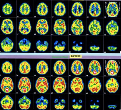
HemoEncephaloGraphy
HEG (hemoencephalography) is the study of blood flow in the brain. More specifically, HEG neurofeedback is the study of voluntarily controlled brain blood flow and corresponding changes (in oxygenation) that are fed back to the user. Hemoencephalography neurofeedback training targets brain areas (usually in the prefrontal cortex) that correspond with particular challenges. For example, the prefrontal cortex (PFC) is involved with executive functions such as attention, organization, and planning. The PFC integrates and analyzes incoming information by way of neuronal connectivity, and mediates other brain area functions with the goal of optimizing survival. Literature reviews reveal that many brain function challenges are rooted in PFC executive function failure. HEG neurofeedback is associated with increased emotional self-regulation (i.e. anger management and bipolar mood swings) by increasing the ability of the PFC to control emotions and reappraise the stimulus.
Before Neurofeedback
After Neurofeedback
HEG neurofeedback allows the trainee to activate their brain which increases blood circulation and thereby strengthens neural connections. Blood delivers oxygen and the basic nutrient glucose (sugar), at life-sustaining levels to the brain. Our brains have an amazing ability to supply extra blood preferentially to areas in current use. Even in repose, the brain consumes about 1/5 of all energy used in the body. Metabolized energy resulting from brain activity causes the brain temperature to rise in the used (venous) blood. If we used the entire brain at one time it would overheat and be severely damaged. The flow of blood cools the brain and prevents overheating.
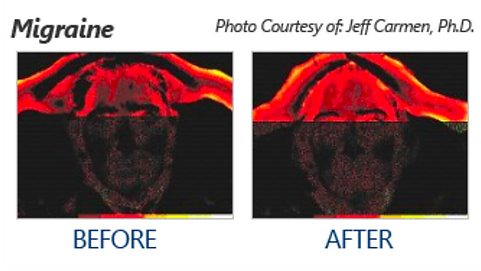
Active brain areas are marked by high oxygen density and higher-than-normal temperatures. Simple measurements can locate active brain areas. Note here that we have no sense that tells us where an active area is located. For example, when we stand, activity occurs in the motor strip across the top of the head, but we have no sensation with information about the exact location of that active bit of brain tissue.
From infancy on, we learn to use specific parts of the brain for accomplishing familiar tasks. We carry out the same learning process for anything new. We may want to do something we see others do, or we try to do something that seems possible. Initially, we accomplish it by trial and error. In trying, we find some modicum of success. We are encouraged, so we try again and again. We improve as we go, and our brains reinforce and expand existing neural networks.
PirHEG (passive infrared) neurofeedback (developed by Jeffrey A. Carmen) uses a sophisticated passive infrared thermometer, and brain temperature changes are fed back to the trainee. nirHEG (near-infrared) neurofeedback uses an optical probe, developed by the originator of HEG Hershel Toomim. nirHEG shines light through the skin and skull to assess the color of brain tissue; oxygenated arterial blood is red, deoxygenated venous blood is blue. Increases in brain activation and demand for nutrition result in faster blood flow and redder blood in the tissues. nirHEG is the study of the change in color of the brain due to brain oxygen absorption. Dr. Toomim, researcher and inventor of nirHEG, described this physiological function as “an outstanding opportunity for providing neurofeedback.”
For more information and related HEG research, please refer to the articles on the Biocomp Research page.